
Abstract
Objective
Systemic inflammation plays a key role in tumor progression and treatment response in advanced non-small cell lung cancer (NSCLC). Among inflammation-based biomarkers, the systemic immune-inflammation index (SII) and systemic inflammatory response index (SIRI) have recently gained attention as potential prognostic tools. This study aimed to evaluate the prognostic impact of SII and SIRI on progression-free survival (PFS) and overall survival (OS) in metastatic NSCLC patients receiving second-line nivolumab.
Method
A retrospective analysis was conducted in 216 patients with metastatic NSCLC who were treated with second-line nivolumab. Baseline hematologic parameters were used to calculate SII and SIRI. Receiver operating characteristic analysis was used to determine optimal cut-off values, and associations between these values and OS and PFS were examined. Clinical variables—including metastatic distribution, response to prior therapy, and nivolumab cycle number—were incorporated into univariate and multivariate Cox regression models.
Results
Median OS and PFS were 27.7 and 12.6 months, respectively, with a median follow-up of 24 months. A SIRI cut-off of ≥2086 was strongly associated with increased mortality and progression risk, while SII demonstrated no significant discriminatory value. An association was observed between the number of nivolumab cycles and survival outcomes, with shorter survival among patients who received fewer cycles. Additionally, the presence of multiorgan metastases and disease progression during prior treatment independently predicted worse outcomes.
Conclusion
SIRI and the nivolumab cycle count appear to be clinically relevant parameters associated with survival outcomes. The lack of prognostic significance for SII suggests that SIRI may be a more reliable inflammation-based marker in this treatment setting. These findings highlight the potential of integrating inflammatory indices with clinical parameters to refine risk stratification and optimize patient management.
Keywords: Nivolumab, non-small cell lung cancer, overall survival, progression-free survival, prognostic biomarkers, systemic immune-inflammation index, systemic inflammatory response index
Introduction
Cancer, excluding non-melanoma skin cancers, remains the most frequently diagnosed malignancy worldwide and represents a major public health problem in terms of both incidence and mortality (). Non-small cell lung cancer (NSCLC) accounts for approximately 85% of all lung cancers and is associated with poor overall survival (OS), largely due to diagnosis at advanced stages and the limited availability of effective treatment options (, ). Because most patients are diagnosed at an advanced stage, survival outcomes remain suboptimal despite current therapeutic strategies (). In recent years, the introduction of immune checkpoint inhibitors (ICIs), particularly anti-PD-1/PD-L1 agents, has marked a major breakthrough in the management of advanced NSCLC and has led to significant improvements in survival outcomes (). However, the response to immunotherapy (IT) varies substantially among patients, highlighting the need for reliable and easily accessible biomarkers that can predict treatment outcomes ().
With increasing understanding of the role of systemic inflammation in tumor development, progression, and metastasis, inflammatory indices derived from peripheral blood parameters have attracted increasing attention (). Among these, the neutrophil-to-lymphocyte ratio (NLR), platelet-to-lymphocyte ratio (PLR), systemic immune-inflammation index (SII), and systemic inflammatory response index (SIRI) are the most commonly investigated biomarkers (). Large-scale meta-analyses in NSCLC populations have demonstrated that elevated NLR levels are significantly associated with poor survival (). Similarly, C-reactive protein (CRP)–based inflammatory scores have been shown to have strong prognostic value across large patient cohorts ().
SIRI is a recently defined index calculated as (neutrophils×monocytes)/lymphocytes (). Meta-analyses including more than 3.000 patients with NSCLC have shown that elevated SIRI levels are consistently associated with poor OS and progression-free survival (PFS) (). Moreover, this association has been reported to remain consistent regardless of differences in country, tumor stage, histology, or cut-off values ().
Recent findings also suggest that SIRI and SII may serve as potential biomarkers for predicting treatment response among patients with advanced NSCLC receiving IT (). However, most existing meta-analyses involve heterogeneous patient populations and combine different treatment modalities, limiting the generalizability of their conclusions. Data specifically evaluating the prognostic roles of SIRI and SII in patients with metastatic NSCLC treated with second-line nivolumab remain limited (). Furthermore, comprehensive studies assessing these inflammatory indices together with clinical features such as metastatic patterns, prior treatment response, and the number of nivolumab cycles are still needed ().
In this study, we aimed to evaluate the prognostic significance of SIRI and SII for OS and PFS in metastatic NSCLC patients receiving second-line nivolumab, as well as their associations with metastatic distribution, number of nivolumab cycles administered, and prior treatment response ().
Materials and Methods
This retrospective study included patients with metastatic NSCLC who received second-line nivolumab treatment at the İstanbul Medipol University Medical Oncology Clinic. Ethics committee approval: Permission was obtained from the İstanbul Medipol University Non-Interventional Clinical Research Ethics Committee (decision no: 1238, date: 16.10.2025). Electronic medical records and radiological data of eligible patients were reviewed.
Patients aged ≥18 years, histologically diagnosed with NSCLC, who had received at least one cycle of nivolumab as second-line therapy and who had accessible baseline laboratory parameters and radiological assessments were included. Patients with insufficient laboratory data, active infections, autoimmune diseases requiring immunosuppressive therapy, hematologic malignancies, or missing follow-up information were excluded.
Demographic characteristics (age, sex), Eastern Cooperative Oncology Group (ECOG) performance status, histopathological subtype, primary tumor location, initial clinical stage, comorbidities, prior treatments, metastatic sites, metastatic tumor burden, number of nivolumab cycles, and radiological treatment responses were obtained from medical records. Pre-treatment neutrophil, lymphocyte, monocyte, and platelet counts, as well as CRP and albumin levels, were retrieved from the hospital laboratory information system. Hemogram analyses were performed on the Mindray CAL 8000 analyzer (Shanghai, China) using electrical-impedance and optical methods, while CRP and albumin levels were measured on Cobas 702 analyzers (Roche Diagnostics, Mannheim, Germany) using the electrochemiluminescence method.
Systemic inflammation indices were calculated using the following formulas:
SII = platelet count × neutrophil count/lymphocyte count
SIRI = neutrophil count × monocyte count/lymphocyte count.
OS was defined as the time from nivolumab initiation until death from any cause. PFS was defined as the time from treatment initiation until radiologically confirmed disease progression or death. Treatment responses were evaluated according to RECIST 1.1 criteria.
Statistical Analysis
Statistical analyses were conducted using IBM SPSS Statistics for Windows, Version 25.0 (IBM Corp., Armonk, NY, USA). The distribution of continuous variables was evaluated using the Kolmogorov-Smirnov test. Normally distributed variables were presented as mean ± standard deviation, whereas non-normally distributed variables were expressed as median (minimum-maximum). Categorical variables were summarized as frequencies and percentages.
Comparisons between groups were performed using the chi-square or Fisher’s exact test for categorical variables, and the independent samples t-test or Mann-Whitney U test for continuous variables. OS and PFS were analyzed using the Kaplan-Meier method, and survival curves were compared using the log-rank test. Prognostic factors affecting survival were examined using Cox proportional hazards regression analysis; hazard ratios (HRs) and 95% confidence intervals (CIs) were reported.
The predictive performance of continuous variables was assessed using receiver operating characteristic (ROC) curve analysis, and optimal cut-off values were determined by the Youden index. The number of nivolumab cycles was included in the analyses as a treatment exposure-related variable, defined as the total number of administered cycles during follow-up. In this study, ≥2086 was used as the cut-off value for SIRI, and ≤10.5 for the number of nivolumab cycles. A p-value <0.05 was considered statistically significant.
To address the potential risk of immortal time bias associated with treatment duration, a landmark analysis was performed for progression-free survival. The landmark time point was defined at 3 months following nivolumab initiation, corresponding to the first routine radiological response assessment. Only patients who were alive and progression-free at the landmark time point were included in the landmark-adjusted multivariate Cox regression model. OS analyses were performed using the full cohort, as the predefined landmark did not result in exclusion of patients or alteration of the OS risk set.
Results
In the present study, 216 patients were included (). The mean age of the patients was 62.56±8.53 years, with a median age of 62.5 (range: 24-88). Among all cases, 64.4% were older than 60 years, and 35.6% were younger than 60 years. The gender distribution revealed that 86.1% of the patients were male and 13.9% were female, indicating a clear predominance of male patients. Regarding performance status, 13.5% of the patients had ECOG 0, 84.2% had ECOG 1, and 2.3% had ECOG 2.
Table 1. Distribution of socio-demographic and clinical variables
Variables | n | % |
|---|---|---|
Age | - | |
Mean ± SD | 62.56±8.53 | |
Median (min-max) | 62.5 (24-88) | |
<60 | 77 | 35.6 |
>60 | 139 | 64.4 |
Sex | - | - |
Female | 30 | 13.9 |
Male | 186 | 86.1 |
ECOG | - | - |
0 | 18 | 13.5 |
1 | 112 | 84.2 |
2 | 3 | 2.3 |
Comorbidity | - | - |
Absent | 95 | 44.0 |
COPD | 34 | 15.7 |
CHF-CAD | 25 | 11.6 |
History of an other malignancy in remission | 9 | 4.2 |
DM or HT | 48 | 22.2 |
Other | 5 | 2.3 |
Histopathology | - | - |
ADC | 99 | 45.8 |
SCC | 81 | 37.5 |
Mixed type | 31 | 14.4 |
Other | 5 | 2.3 |
Primary tumor location | - | - |
Right upper lobe | 83 | 38.4 |
Right middle lobe | 17 | 7.9 |
Right lower lobe | 31 | 14.4 |
Left upper lobe | 41 | 19.0 |
Left lower lobe | 29 | 13.4 |
Central | 15 | 6.9 |
Initial stage | - | - |
Early stage at diagnosis | 99 | 45.8 |
Metastatic at diagnosis | 117 | 54.2 |
Vascular invasion | - | - |
Unknown | 175 | 81.0 |
Present | 19 | 8.8 |
Absent | 22 | 10.2 |
Lymphatic invasion | - | - |
Unknown | 175 | 81.0 |
Present | 19 | 8.8 |
Absent | 22 | 10.2 |
Perineural invasion | - | - |
Unknown | 172 | 79.6 |
Present | 14 | 6.5 |
Absent | 30 | 13.9 |
RT type | - | - |
Received adjuvant RT | 1 | 1.9 |
Definitive CRT | 44 | 84.6 |
Sequential CT and RT | 7 | 13.5 |
RT with concurrent CT | - | - |
No concurrent chemotherapy | 7 | 13.5 |
Concurrent carboplatin–paclitaxel | 44 | 84.6 |
Concurrent chemotherapy unknown | 1 | 1.9 |
Metastasis sites | - | - |
Lung widespread | 50 | 23.1 |
Lung local | 34 | 15.7 |
Brain metastasis | 24 | 11.1 |
Bone metastasis | 27 | 12.5 |
Multiorgan | 77 | 35.6 |
Adrenal | 4 | 1.9 |
Tumor burden in metastatic disease | - | - |
Oligometastatic 1-2-3 lesions | 72 | 33.3 |
Oligometastatic 4-5 lesions | 39 | 18.1 |
No Oligometastatic disease | 75 | 34.7 |
Local-regional recurrence only | 30 | 13.9 |
Initial treatment summary | - | - |
Induction CT then CRT | 16 | 7.4 |
CRT then consolidation CT | 12 | 5.6 |
Received sequential RT/CT | 5 | 2.3 |
Previously operated + adjuvant CT | 25 | 11.6 |
Previously operated + adjuvant RT/CT | 6 | 2.8 |
Follow-up after definitive CRT | 31 | 14.4 |
Postoperative follow-up, recurrence treated with RT/CT | 8 | 3.7 |
Nivolumab cycle count | - | - |
Mean ± SD | 13.96±11.07 | - |
Median (min-max) | 10.00 (1.00-50.00) | - |
SUVmax on PET before nivolumab | - | - |
Mean ± SD | 12.65±6.85 | - |
Median (min-max) | 11.30 (2.80-42.80) | - |
Received RT during immunotherapy | - | - |
No | 184 | 85.2 |
Yes | 32 | 14.8 |
Radiologic interim response to immunotherapy | - | - |
Complete response | 11 | 5.3 |
Partial response | 64 | 30.8 |
Stable disease | 43 | 20.7 |
Unconfirmed progression | 4 | 1.9 |
Confirmed progression | 84 | 40.4 |
Pseudoprogression | 2 | 1.0 |
Recurrence sites after immunotherapy | - | - |
Absent | 62 | 32.8 |
Lung local | 27 | 14.3 |
Brain metastasis | 10 | 5.3 |
Bone metastasis | 9 | 4.8 |
Liver metastasis | 1 | 0.5 |
Multiorgan | 80 | 42.3 |
Relationship to prior treatment | - | - |
Progressed during prior treatment | 154 | 71.3 |
Progressed during drug-free follow-up | 62 | 28.7 |
Chemotherapy response before immunotherapy | - | - |
Complete response | 8 | 3.7 |
Partial response | 55 | 25.5 |
Stable response | 20 | 9.3 |
Progressive disease | 127 | 58.8 |
Unknown | 6 | 2.8 |
SII | - | - |
Mean ± SD | 286219.00±110497.80 | - |
Median (min-max) | 260847.0 (21995.0-736000.0) | - |
SIRI | - | - |
Mean ± SD | 3002.84±2847.96 | - |
Median (min-max) | 2086.00 (175.0-21144.0) | - |
Progression | - | - |
No | 91 | 42.1 |
Yes | 125 | 57.9 |
Mortality | - | - |
Alive | 107 | 49.5 |
Dead | 109 | 50.5 |
Follow-up duration (months) | - | - |
Mean ± SD | 24.43±17.28 | - |
Median (min-max) | 20.16 (1.03-117.20) | - |
SD: Standard deviation, ADC: Adenocarcinoma, SCC: Squamous cell carcinoma, CHF: Congestive heart failure, CAD: Coronary artery disease, COPD: Chronic obstructive pulmonary disease, DM: Diabetes mellitus, HT: Hypertension, ECOG: Eastern Cooperative Oncology Group, SIRI: Systemic inflammatory response index, SII: Systemic immune-inflammation index, PET: Positron emission tomography, CRT: Chemoradiotherapy, RT: Radiotherapy, CT: Computed tomography
Regarding comorbidities, 44.0% of patients had none. The most common comorbidities were diabetes mellitus or hypertension (22.2%), COPD (15.7%), and heart failure-coronary artery disease (11.6%). A history of another malignancy in remission was reported in 4.2% of patients, whereas 2.3% had other less frequent comorbidities.
Histopathological evaluation showed that 45.8% of cases had adenocarcinoma, 37.5% had squamous cell carcinoma, 14.4% had mixed type, and 2.3% had other histological subtypes. Regarding primary tumor localization, the most common site was the right upper lobe (38.4%), followed by the left upper lobe (19.0%), the right lower lobe (14.4%), the left lower lobe (13.4%), and the right middle lobe (7.9%). Centrally located tumors accounted for 6.9% of all cases.
At diagnosis, 54.2% of the patients presented with metastatic disease, while 45.8% were diagnosed at an early stage. Vascular, lymphatic, and perineural invasion rates were 8.8%, 8.8%, and 6.5%, respectively.
Regarding radiotherapy, the majority of patients (84.6%) underwent definitive chemoradiotherapy. The most commonly administered regimen among those receiving concurrent chemotherapy was carboplatin-paclitaxel (84.6%); 13.5% did not receive concurrent chemotherapy.
Assessment of metastatic involvement revealed that 23.1% of patients had diffuse lung metastases, 15.7% had localized lung metastases, 11.1% had brain metastases, 12.5% had bone metastases, 1.9% had adrenal metastases, and 35.6% had multiple-organ metastatic involvement. Based on metastatic tumor burden, 33.3% were classified as oligometastatic with 1-3 lesions, 18.1% with 4-5 lesions, 34.7% had no oligometastatic disease, and 13.9% had locoregional recurrence only.
Evaluation of initial treatment strategies demonstrated that 52.3% of patients presented with metastatic disease at diagnosis and received systemic therapy. Additionally, 11.6% underwent surgery followed by adjuvant chemotherapy; 14.4% were monitored after definitive chemoradiotherapy; 7.4% received induction chemotherapy followed by chemoradiotherapy; 5.6% received consolidation chemotherapy following chemoradiotherapy; 2.3% received sequential radiotherapy/chemotherapy; and 3.7% were treated with radiotherapy/chemotherapy for recurrence after surgery.
Regarding IT, patients received a mean of 13.96±11.07 cycles of nivolumab (median: 10 cycles; range: 1-50). The mean pre‑IT baseline PET SUVmax value was 12.65±6.85 (median: 11.30; range: 2.80-42.80). Radiotherapy during IT was administered to 14.8% of patients.
Radiological response analysis showed complete response in 5.3% of patients, partial response in 30.8%, stable disease in 20.7%, confirmed progression in 40.4%, and pseudoprogression in 1.0%. Following IT, multiorgan recurrence was the most common relapse pattern (42.3%), followed by localized lung (14.3%), brain (5.3%), bone (4.8%), and liver (0.5%) recurrences; no recurrence was observed in 32.8% of patients.
Regarding pre‑IT treatment status, 71.3% of patients had progressed during prior therapy, while 28.7% experienced progression during treatment‑free follow‑up. Pre‑IT chemotherapy response assessment showed complete response in 3.7%, partial response in 25.5%, stable disease in 9.3%, and progressive disease in 58.8% of patients.
Evaluation of systemic inflammatory markers revealed a mean SII of 286219.00±110497.80 and a mean SIRI of 3002.84±2847.96. The mean follow‑up duration was 24.43±17.28 months (median: 20.16; range: 1.03-117.20 months). The progression rate was 57.9%, and the mortality rate was 50.5%.
As shown in , the predictive value of various clinical parameters for distinguishing mortality was evaluated using ROC analysis. According to the results, the number of nivolumab cycles demonstrated discriminative ability with respect to mortality [area under the curve (AUC)=0.843; 95% confidence interval (CI): 0.792-0.893; p<0.001]. The determined cut-off value was ≤10.50, indicating that patients receiving nivolumab below this threshold had a significantly higher risk of mortality. Sensitivity and specificity at this cut-off were calculated as 77.1% and 76.4%, respectively.
Table 2. Analysis of the predictive value of various clinical parameters in distinguishing mortality
Variables | AUC | 95% CI | Cut-off | Sensitivity (%) | Specificity (%) | p |
SII | 0.554 | 0.477-0.631 | ≥260847.00 | 55.0 | 55.2 | 0.173 |
SIRI | 0.652 | 0.578-0.725 | ≥2086.00 | 60.6 | 61.0 | <0.001 |
Number of nivolumab cycles | 0.843 | 0.792-0.893 | ≤10.50 | 77.1 | 76.4 | <0.001 |
AUC: Area under the curve, CI: Confidence interval, SIRI: Systemic inflammatory response index, SII: Systemic immune-inflammation index
SIRI was also found to be a significant predictor of mortality (AUC=0.652; 95% CI: 0.578-0.725; p<0.001). The cut-off value of ≥2086.00 was identified as the threshold associated with increased mortality risk, with sensitivity of 60.6% and specificity of 61.0%.
In contrast, SII did not demonstrate a statistically significant discriminative ability for mortality (AUC=0.554; 95% CI: 0.477-0.631; p=0.173).
As shown in , the median OS for all patients was 27.66 months (95% CI: 19.31-36.01). Median OS differed significantly according to several clinical variables, including initial disease stage (p=0.001), metastatic sites (p=0.008), initial treatment summary (p=0.034), number of nivolumab cycles (p<0.001), the relationship between prior treatment and IT (p=0.002), chemotherapy response before initiation of IT (p=0.004), and SIRI groups (p<0.001) (, , , ).
Table 3. OS comparisons of the patients
Variables | 2-year % | 5-year % | Median months (95% CI) | p |
|---|---|---|---|---|
Overall | 56.8 | 37.1 | 27.66 (19.31-36.01) | - |
Age | - | - | - | - |
<60 | 60.0 | 35.7 | 34.80 (11.92-57.67) | 0.478 |
>60 | 55.2 | 36.8 | 27.16 (17.67-36.66) | |
Sex | - | - | - | - |
Female | 54.0 | - | 26.33 (15.20-37.45) | 0.941 |
Male | 57.1 | 37.1 | 27.66 (19.17-36.15) | |
Initial stage | - | - | - | - |
Early stage at diagnosis | 67.9 | 44.2 | 43.46 (18.60-68.32) | 0.001 |
Metastatic at diagnosis | 43.8 | 35.3 | 22.13 (18.75-25.50) | |
Lymphatic invasion | - | - | - | - |
Present | 69.4 | 39.2 | 29.66 (6.39-52.93) | 0.766 |
Absent | 61.3 | 51.1 | - (-) | |
PNI | - | - | - | - |
Present | 64.3 | 48.2 | 27.16 (0.0-73.79) | 0.966 |
Absent | 68.8 | 41.6 | 43.46 (34.50-52.42) | |
RT type | - | - | - | - |
Definitive CRT | 60.9 | 36.9 | 36.33 (7.83-64.83) | 0.213 |
Sequential CT and RT | 42.9 | - | 24.00 (11.51-36.48) | |
Metastasis sites | - | - | - | - |
Lung widespread | 74.3 | 58.1 | 67.73 (0.00-140.77) | 0.008 |
Lung local | 7.5 | 46.4 | 60.00 (25.62-94.37) | |
Brain metastasis | 46.0 | - | 23.66 (17.78-29.54) | |
Bone metastasis | 43.6 | 43.6 | 20.20 (18.40-21.99) | |
Multiorgan | 44.4 | 27.1 | 19.63 (14.77-24.48) | |
Tumor burden in metastatic disease | - | - | - | - |
Oligometastatic 1-2-3 lesions | 57.4 | 38.7 | 34.80 (18.12-51.47) | 0.178 |
Oligometastatic 4-5 lesions | 58.6 | 37.5 | 36.33 (14.62-58.04) | |
No Oligometastatic disease | 46.1 | 32.3 | 20.96 (13.33-28.59) | |
Local-regional recurrence only | 76.4 | 47.8 | 60.00 (20.63-99.36) | |
Initial treatment summary | - | - | - | - |
Induction CT then CRT | 61.9 | 37.7 | 26.33 (24.05-28.61) | 0.034 |
CRT then consolidation CT | 43.8 | 32.8 | 23.66 (17.05-30.28) | |
Received sequential RT/CT | 26.7 | - | 24.00 (17.05-30.94) | |
Previously operated + adjuvant CT | 79.6 | 56.5 | 60.40 (20.98-99.81) | |
Metastatic at diagnosis | 46.0 | 34.8 | 22.13 (18.45-25.80) | |
Previously operated + adjuvant RT/CT | 83.3 | 62.5 | 78.23 (-) | |
Follow-up after definitive CRT | 71.0 | 42.4 | 60.00 (22.74-97.25) | |
Post-op follow-up, recurrence treated with RT/CT | 62.5 | 31.3 | 43.46 (10.02-76.90) | |
Nivolumab cycle count | - | - | - | - |
>10.50 | 83.9 | 64.9 | 117.20 (-) | <0.001 |
≤10.50 | 30.2 | 14.1 | 15.63 (12.59-18.67) | |
Relationship to prior treatment | - | - | - | - |
Progressed during prior treatment | 49.3 | 29.4 | 24.00 (16.38-31.61) | 0.002 |
Progressed during drug-free follow-up | 74.3 | 52.7 | 60.40 (11.52-109.27) | |
Chemotherapy response before immunotherapy | - | - | - | - |
Complete response | 87.5 | 87.5 | 60.40 (13.85-106.94) | 0.004 |
Partial response | 68.7 | 42.9 | 29.93 (18.06-41.79) | |
Stable response | 69.6 | 58.0 | - (-) | |
Progressive disease | 44.9 | 26.8 | 20.96 (16.36-25.56) | |
SIRI | - | - | - | - |
<2086.00 | 71.9 | 46.6 | 60.00 (31.70-88.29) | <0.001 |
≥2086.00 | 41.5 | 26.6 | 19.63 (15.09-24.17) |
Kaplan-Meier analysis and log-rank test were applied; p<0.05 was considered statistically significant, SIRI: Systemic inflammatory response index, RT: Radiotherapy, CRT: Chemoradiotherapy, CT: Computed tomography, OS: Overall survival, CI: Confidence interval, PNI: Perineural invasion
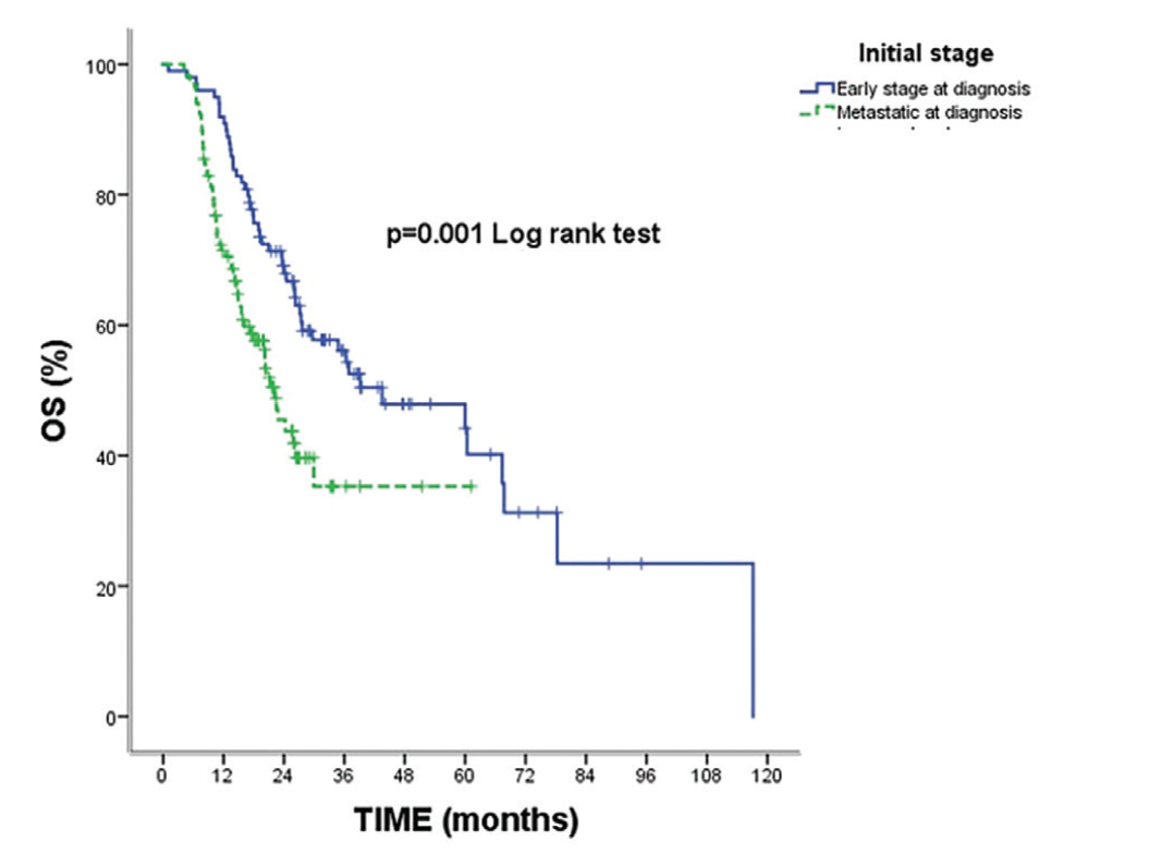
SIRI: Systemic inflammatory response index
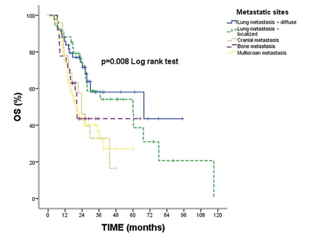
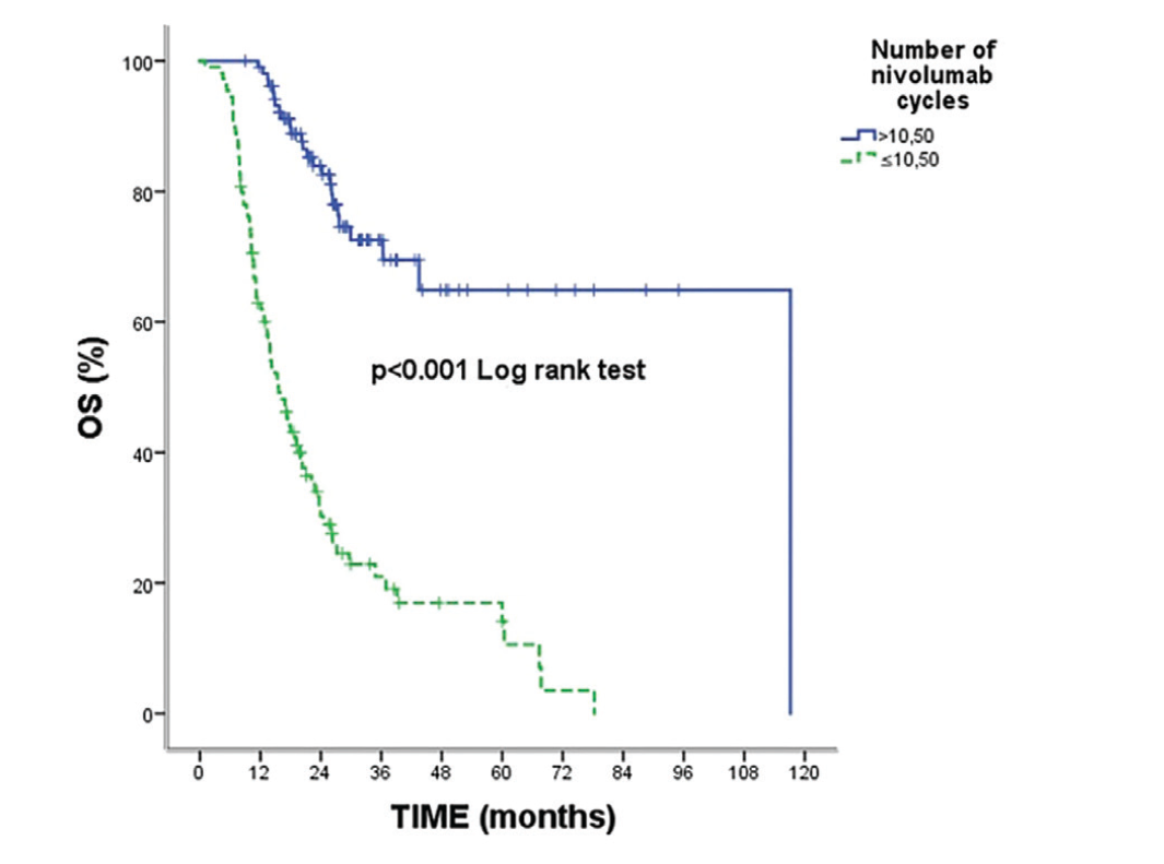
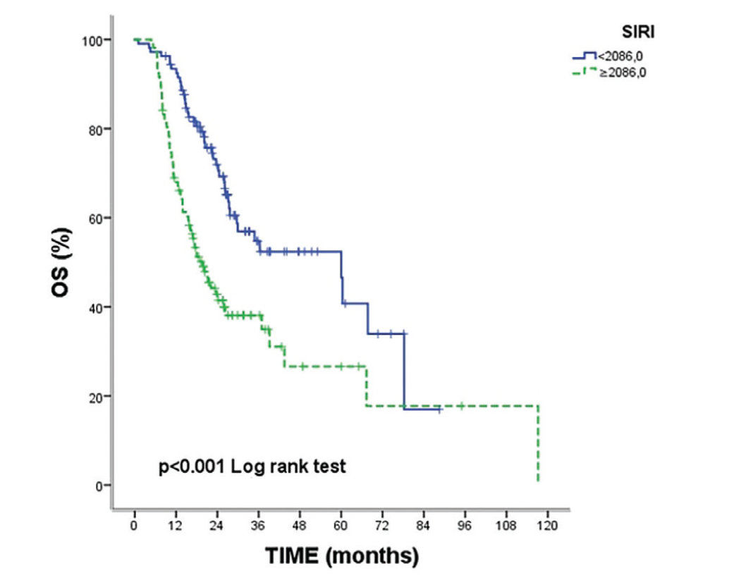
SIRI: Systemic inflammatory response index
As shown in , the overall median PFS was 12.56 months (95% CI: 9.09-16.03). Median PFS differed significantly by metastasis site (p=0.026), number of nivolumab cycles (p<0.001), the relationship between prior treatment and IT (p=0.001), and SIRI group (p<0.001). Patients with multiorgan metastasis and those who received ≤10.50 nivolumab cycles had markedly shorter PFS (, , , ).
Table 4. PFS comparisons of the patients
Variables | 2-year % | 5-year % | Median months (95% CI) | p |
|---|---|---|---|---|
Overall | 37.0 | 13.9 | 12.56 (9.09-16.03) | - |
Age | - | - | - | - |
<60 | 46.2 | - | 19.86 (11.73-27.99) | 0.054 |
>60 | 32.6 | 20.7 | 11.36 (8.81-13.91) | |
Sex | - | - | - | - |
Female | 34.3 | - | 13.50 (8.49-18.50) | 0.899 |
Male | 37.3 | 13.3 | 12.56 (8.69-16.43) | |
Initial stage | - | - | - | - |
Early stage at diagnosis | 39.9 | 27.6 | 18.16 (13.32-23.01) | 0.056 |
Metastatic at diagnosis | 37.1 | - | 9.90 (7.90-11.80) | |
Lymphatic invasion | - | - | - | - |
Present | 31.4 | - | 18.16 (6.85-29.47) | 0.851 |
Absent | 45.9 | - | 12.66 (1.80-23.52) | |
PNI | - | - | - | - |
Present | 45.1 | - | 16.16 (-) | 0.788 |
Absent | 34.7 | - | 18.16 (8.07-28.25) | |
RT type | - | - | - | - |
Definitive CRT | 43.3 | 11.9 | 15.56 (7.67-23.46) | 0.074 |
Sequential CT and RT | - | - | 9.86 (8.92-10.80) | |
Metastasis sites | - | - | - | - |
Lung widespread | 54.4 | - | 25.53 (-) | 0.026 |
Lung local | 44.9 | 22.5 | 15.56 (8.56-22.56) | |
Brain metastasis | 20.6 | - | 16.16 (5.69-26.63) | |
Bone metastasis | 42.3 | - | 11.13 (7.61-14.65) | |
Multiorgan | 26.4 | - | 8.06 (5.64-10.48) | |
Tumor burden in metastatic disease | - | - | - | - |
Oligometastatic 1-2-3 lesions | 40.9 | 14.7 | 17.56 (9.90-25.22) | 0.065 |
Oligometastatic 4-5 lesions | 29.3 | - | 7.93 (2.01-13.86) | |
No Oligometastatic disease | 31.2 | - | 9.86 (5.27-14.46) | |
Local-regional recurrence only | 51.7 | - | 25.03 (-) | |
Initial treatment summary | - | - | - | - |
Induction CT then CRT | 36.5 | - | 11.73 (7.48-15.98) | 0.179 |
CRT then consolidation CT | 31.3 | - | 11.13 (7.96-14.30) | |
Received sequential RT/CT | - | - | 9.50 (7.78-11.21) | |
Previously operated + adjuvant CT | 48.4 | - | 19.86 (-) | |
Metastatic at diagnosis | 35.9 | - | 9.66 (7.15-12.18) | |
Previously operated + adjuvant RT/CT | 50.0 | - | 6.56 (-) | |
Follow-up after definitive CRT | 49.7 | 19.0 | 18.73 (7.73-29.73) | |
Post-op follow-up, recurrence treated with RT/CT | 34.3 | - | 21.43 (0.00-45.53) | |
Nivolumab cycle count | ||||
>10.50 | 60.5 | 23.5 | 35.63 (24.25-47.01) | <0.001 |
≤10.50 | 10.4 | - | 5.80 (5.20-6.39) | |
Relationship to prior treatment | - | - | - | - |
Progressed during prior treatment | 33.4 | - | 9.13 (6.64-11.62) | 0.001 |
Progressed during drug-free follow-up | 48.6 | 16.5 | 19.86 (13.03-26.69) | |
Chemotherapy response before immunotherapy | - | - | - | - |
Complete response | 75.0 | - | 25.60 (15.68-35.52) | 0.162 |
Partial response | 34.8 | 27.8 | 15.56 (10.96-20.17) | |
Stable response | 40.2 | - | 11.73 (8.14-15.32) | |
Progressive disease | 36.3 | - | 9.13 (6.15-12.10) | |
SIRI | - | - | - | - |
<2086.00 | 47.0 | 17.8 | 18.73 (10.62-26.84) | <0.001 |
≥2086.00 | 17.3 | - | 8.76 (6.34-11.18) |
Kaplan-Meier survival analysis and log-rank test were applied; p<0.05 was accepted as statistically significant, PFS: Progression-free survival, CT: Computed tomography, RT: Radiotherapy, CRT: Chemoradiotherapy, SIRI: Systemic inflammatory response index, PNI: Perineural invasion, CI: Confidence interval
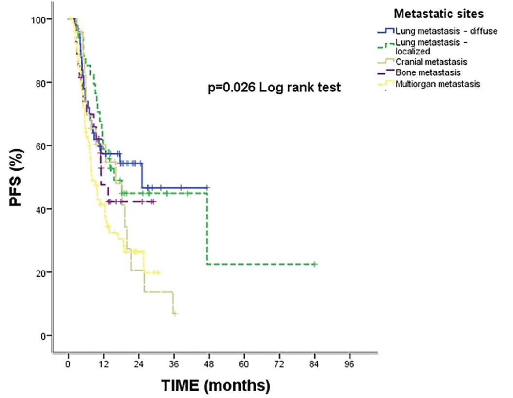
SIRI: Systemic inflammatory response index
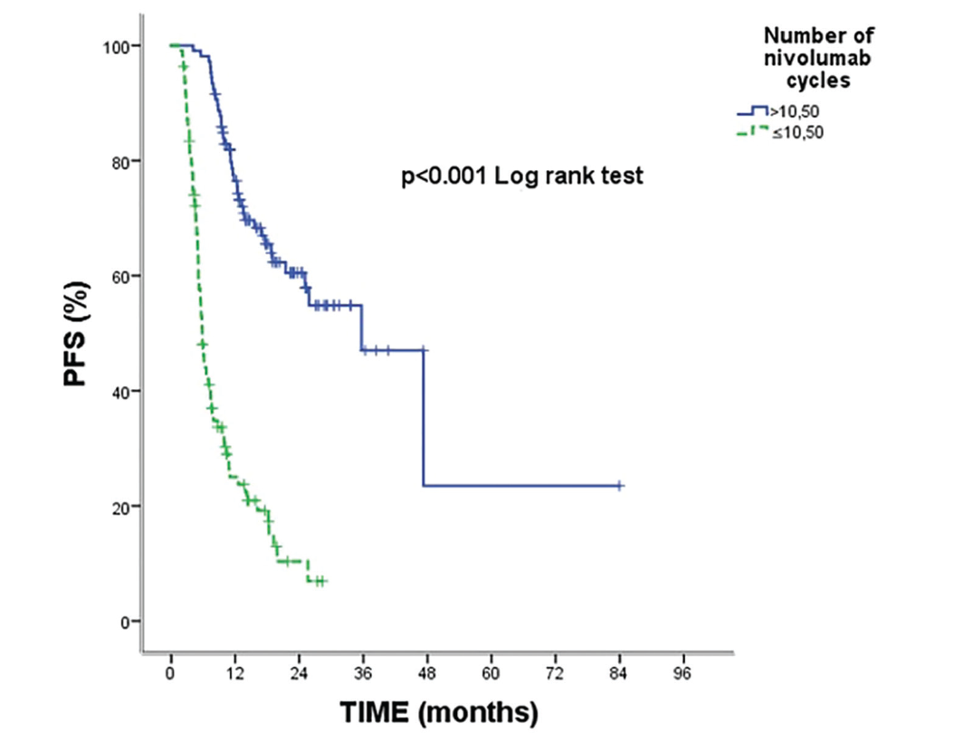
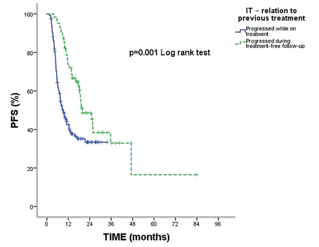
IT: Immunotherapy
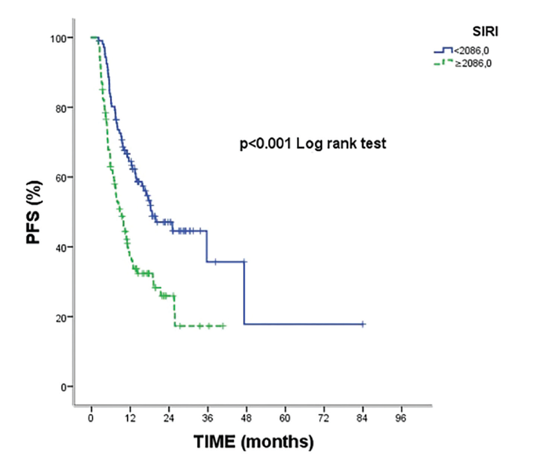
SIRI: Systemic inflammatory response index
As shown in , the variables initial stage, metastasis sites, initial treatment summary, number of nivolumab cycles, relationship to prior treatment, chemotherapy response before IT, and SIRI were found to be significant in the univariate analyses. Variables identified as significant in the univariate analyses were included in the multivariate Cox regression model. According to the model results, being in the lung local group increased the risk of death by 2.29-fold (HR: 2.29, 95% CI: 1.01-5.24, p=0.050), having brain metastases by 2.67-fold (HR: 2.67, 95% CI: 1.19-5.98, p=0.017), having bone metastasis by 2.45-fold (HR: 2.45, 95% CI: 1.15-5.21, p=0.019), having multiorgan metastasis by 2.69-fold (HR: 2.69, 95% CI: 1.45-5.02, p=0.002), having ≤10.50 nivolumab cycles by 6.38-fold (HR: 6.38, 95% CI: 3.90-10.43, p<0.001), and having progressive disease by 5.24-fold (HR: 5.24, 95% CI: 1.09-25.07, p=0.038) were determined to increase the risk of death.
Table 5. Multivariate Cox regression results of various clinical variables on mortality risk
Variables | HR (95% CI) | p |
|---|---|---|
Initial stage | - | - |
Early stage at diagnosis | ref | 0.249 |
Metastatic at diagnosis | 2.40 (0.54-10.69) | |
Metastasis sites | - | 0.032 |
Lung widespread | ref | - |
Lung local | 2.29 (1.01-5.24) | 0.050 |
Brain metastasis | 2.67 (1.19-5.98) | 0.017 |
Bone metastasis | 2.45 (1.15-5.21) | 0.019 |
Multiorgan | 2.69 (1.45-5.02) | 0.002 |
Initial treatment summary | - | 0.381 |
Induction CT then CRT | ref | - |
CRT then consolidation CT | 0.69 (0.20-1.70) | 0.333 |
Received sequential RT/CT | 0.63 (0.16-2.47) | 0.512 |
Previously operated + adjuvant CT | 0.29 (0.10-0.89) | 0.055 |
Metastatic at diagnosis | 0.52 (0.21-1.29) | 0.633 |
Previously operated + adjuvant RT/CT | 0.19 (0.04-0.88) | 0.065 |
Follow-up after definitive CRT | 0.52 (0.21-1.29) | 0.161 |
Post-op follow-up, recurrence treated with RT/CT | 0.53 (0.16-1.71) | 0.294 |
Nivolumab cycle count | - | - |
>10.50 | ref | <0.001 |
≤10.50 | 6.38 (3.90-10.43) | |
Relationship to prior treatment | - | - |
Progressed during prior treatment | ref | 0.400 |
Progressed during drug-free follow-up | 1.35 (0.66-2.77) | |
Chemotherapy response before immunotherapy | - | 0.018 |
Complete response | ref | - |
Partial response | 2.02 (0.44-9.17) | 0.359 |
Stable response | 4.12 (0.77-21.88) | 0.096 |
Progressive disease | 5.24 (1.09-25.07) | 0.038 |
SIRI | - | - |
<2086.00 | ref | 0.111 |
≥2086.00 | 1.41 (0.92-2.17) |
-2 Log Likelihood:865.04, p<0.001, SIRI: Systemic inflammatory response index, CT: Computed tomography, RT: Radiotherapy,
CRT: Chemoradiotherapy, HR: Hazard ratio, CI: Confidence interval
As shown in , metastatic sites, number of nivolumab cycles, relationship to prior treatment, and SIRI were found to be significant in the univariate analyses. To minimize potential immortal time bias related to treatment duration, a landmark analysis was performed, and patients who were alive and progression-free at the predefined landmark time point were included in the multivariate Cox regression model. According to the landmark-adjusted model results, having multiorgan metastasis was associated with an increased risk of progression (HR: 1.77, 95% CI: 0.54-2.98, p=0.051), and receiving ≤10.50 nivolumab cycles by the landmark time point was associated with a markedly higher risk of progression (HR: 6.48, 95% CI: 4.13-10.16, p<0.001). In contrast, progression during drug-free follow-up was associated with a reduced risk (HR: 0.30, 95% CI: 0.19-0.49, p<0.001).
Table 6. Landmark-adjusted multivariate Cox regression analysis of clinical variables associated with progression risk
Variables | HR (95% CI) | p |
|---|---|---|
Metastasis sites | - | 0.057 |
Lung widespread | ref | - |
Lung local | 1.13 (0.59-2.17) | 0.709 |
Brain metastasis | 1.42 (0.73-2.76) | 0.298 |
Bone metastasis | 0.73 (0.35-1.51) | 0.401 |
Multiorgan | 1.77 (0.54-2.98) | 0.051 |
Nivolumab cycle count | - | - |
>10.50 | ref | <0.001 |
≤10.50 | 6.48 (4.13-10.16) | |
Relationship to prior treatment | - | - |
Progressed during prior treatment | ref | <0.001 |
Progressed during drug-free follow-up | 0.30 (0.19-0.49) | |
SIRI | - | - |
<2086.00 | ref | 0.280 |
≥2086.00 | 1.24 (0.83-1.83) |
-2 Log Likelihood:1073.11, p<0.001, HR: Hazard ratio, CI: Confidence interval, SIRI: Systemic inflammatory response index
Discussion
In this study, the relationship among SIRI, SII, and prognosis was evaluated using real-world data from patients with metastatic NSCLC treated with second-line nivolumab. With a mean follow-up of 24 months, the median OS and PFS were 27.7 and 12.6 months, respectively. In the ROC analysis, a SIRI cut-off value of ≥2086 was associated with an increased risk of both mortality and progression, whereas SII did not demonstrate similar discriminatory performance. One of the notable findings of our study was that the number of nivolumab cycles was consistently associated with both OS and progression-free survival. This association should be interpreted with caution because treatment duration is closely linked to underlying disease biology and treatment response; patients with better disease control are therefore more likely to receive a greater number of treatment cycles. Therefore, the number of nivolumab cycles should be considered a surrogate marker of clinical course rather than an independent causal determinant of survival. In the multivariate analyses, multiorgan metastasis remained an independent adverse prognostic factor, whereas progression during drug-free follow-up was associated with a reduced risk of death.
The relatively long median OS and PFS observed in our cohort, compared with pivotal randomized trials of second-line nivolumab, may be explained by differences in patient selection and disease characteristics. Nearly half of the patients were initially diagnosed at an early stage and received definitive local treatment before recurrence, a subgroup known to have more favorable tumor biology and a lower metastatic burden. In addition, a considerable proportion of patients experienced progression during drug-free follow-up; this progression is generally associated with better prognosis and treatment sensitivity. Therefore, the observed survival outcomes likely reflect real-world population heterogeneity rather than methodological bias.
Systemic inflammation-based indices have strong prognostic value in many solid tumors, as demonstrated consistently in large meta-analyses across various cancer types. Several studies have reported that SIRI, which reflects the combined dynamics of neutrophils, monocytes, and lymphocytes, is an independent poor prognostic factor for both OS and PFS in gastrointestinal, gynecological, and genitourinary malignancies (-). Similarly, meta-analytic findings have demonstrated that SII is associated with high tumor burden, advanced stage, and poor survival, particularly in gastrointestinal system tumors (, ). In our study, the finding that SIRI was significant for both OS and PFS in univariate analyses is consistent with the broader cancer literature and suggests that SIRI may be a sensitive indicator of systemic inflammatory burden in metastatic NSCLC patients receiving IT.
When studies focusing specifically on NSCLC are examined, the prognostic role of peripheral blood inflammation indices becomes even more prominent. Systematic reviews and meta-analyses including patients with advanced lung cancer have shown that elevated SII levels adversely affect both OS and PFS in both early-stage resected patients and metastatic cases, and that SII often demonstrates stronger prognostic performance than traditional parameters such as NLR and PLR (-). Likewise, retrospective cohorts have shown that SIRI is associated with tumor stage, metastatic burden, and survival in NSCLC (). The observation in our cohort that SII did not show significant discriminatory ability in either the ROC analysis or the survival curves is partially inconsistent with the literature. This may be related to the structure of our selected cohort, differences in SII cut-off values, sample size, and the effects of radiotherapy on PLRs (-).
Although both SII and SIRI are derived from peripheral blood inflammatory parameters, they reflect distinct biological aspects of the host–tumor interaction. SIRI incorporates monocytes in addition to neutrophils and lymphocytes, thereby better capturing monocyte-driven immunosuppressive mechanisms that are particularly relevant in the context of immune checkpoint inhibitor therapy (, ). Monocytes and tumor-associated macrophages play pivotal roles in shaping the tumor microenvironment and facilitating immune evasion, which may directly influence the response to nivolumab (). In contrast, SII does not directly account for monocyte-related immune suppression and may be more susceptible to treatment-related fluctuations in platelet and lymphocyte counts, such as those induced by radiotherapy or peri-treatment inflammatory changes (-). These biological and treatment-related differences may partly explain the observed discrepancy between the prognostic performances of SIRI and SII in our cohort.
In recent years, a growing body of evidence suggests that SII and SIRI may serve as useful biomarkers for predicting treatment response and survival in patients with metastatic lung cancer treated with ICIs. Meta-analyses including large patient series have shown that elevated pre-treatment SII levels are associated with significantly worse OS and PFS, and that this relationship is maintained across different tumor types and treatment lines (-). Meta-analyses specific to advanced lung cancer have also reported markedly shortened survival for patients with high baseline SII despite ICI therapy. The lack of significance of SII in our study suggests that SII may be less predictive than expected in a homogeneous subgroup of metastatic NSCLC patients receiving IT, and is consistent with the literature suggesting that dynamic changes in SII may be more meaningful ().
In our univariate analyses, SIRI was found to be significant for both OS and PFS, a finding that is more consistent with the IT literature. Studies including lung cancer patients treated with ICIs have reported that high SIRI levels are associated with low treatment response rates, shorter PFS, and shorter OS (). The ability of SIRI to predict IT response has been proposed to be related to neutrophil- and monocyte-mediated immunosuppression. The loss of significance of SIRI in our multivariate model may be due to its coexistence with other strong prognostic factors, such as nivolumab cycle count and metastatic burden.
One of the key findings of our study was that the number of nivolumab cycles was independently associated with both OS and PFS. The ROC analysis yielded an AUC of 0.84, and patients who received ≤10.5 cycles experienced a markedly higher risk of mortality and progression. However, this association should be interpreted cautiously, as patients with better disease control are more likely to receive a higher number of treatment cycles. In the literature, the optimal duration of immune checkpoint inhibitor therapy and the concept of treatment beyond progression remain controversial, with most available evidence derived from secondary or exploratory analyses. In this context, our real-world data highlight a strong prognostic association between treatment duration and clinical outcomes in patients with metastatic NSCLC receiving second-line nivolumab.
To mitigate the potential risk of immortal time bias related to treatment duration, a landmark analysis was applied to progression-free survival, with the landmark time point defined as 3 months after nivolumab initiation. Only patients who were alive and progression-free at the landmark were included in the landmark-adjusted multivariate Cox regression model. OS analyses were performed using the full cohort, as the predefined landmark did not result in exclusion of patients or alteration of the OS risk set.
Findings regarding patterns of metastasis and tumor burden were consistent with those observed in inflammation-based indices. The finding that multiorgan metastasis was an independent poor prognostic factor for both OS and PFS is consistent with studies reporting higher SII levels in patients with brain and bone metastases (, ). In our study, the inclusion of metastatic burden in the model, independent of SIRI/SII, indicates that inflammation scores alone may not fully explain the prognostic heterogeneity. Therefore, using SIRI and SII together with metastatic burden may provide a more accurate risk stratification.
Treatment response prior to IT and timing of progression are also important prognostic indicators. Poorer survival among patients who progressed on prior therapy reflects biological aggressiveness and reduced sensitivity to treatment. In contrast, longer OS in patients who progressed during drug-free follow-up after chemotherapy is consistent with the concept of “chemo-sensitive disease” (, ). These findings suggest that SIRI and SII should be evaluated together with clinical parameters.
A key clinical contribution of our study is the introduction of a practical basis for risk stratification that combines SIRI with nivolumab cycle count in metastatic NSCLC patients receiving second-line IT. Patients with high SIRI who discontinue nivolumab early may be defined as a high-risk group, whereas those with low SIRI receiving prolonged nivolumab therapy may represent a subgroup likely to derive long-term benefit (, ). Validation of this approach in prospective studies may contribute to the development of risk-based treatment and follow-up algorithms in real-world practice.
Study Limitations
This study has several limitations, including its retrospective design, single-center setting, heterogeneity of cut-off values, lack of biomarker data, such as PD-L1 and TMB, and the assessment of SIRI and SII only at baseline. Literature suggests that dynamic measurements may provide greater prognostic insight (). Although a landmark approach was applied to mitigate immortal time bias, residual confounding related to treatment duration cannot be completely excluded.
Conclusion
In conclusion, in this real-world cohort of patients with metastatic NSCLC treated with second-line nivolumab, SIRI demonstrated significant prognostic value for both OS and progression-free survival. Although SII did not show discriminatory performance, elevated SIRI levels were associated with poorer outcomes. The strongest prognostic factor identified in the study was the number of nivolumab cycles received; patients who discontinued treatment early (≤10.5 cycles) had a markedly higher risk of both mortality and progression. In addition, multiorgan metastatic disease was confirmed as an independent adverse prognostic factor.
These findings highlight the clinical utility of SIRI and treatment duration as practical, easily accessible parameters for risk stratification in patients receiving ICIs. Combining systemic inflammation markers with treatment-related variables, such as nivolumab cycle count, may facilitate more individualized prognostic assessment. Prospective studies evaluating dynamic changes in inflammatory indices and integrating additional biomarkers, including PD-L1 and TMB, are needed to further refine prognostic models and guide treatment optimization in metastatic NSCLC.
Footnotes
Ethics
Ethics Committee Approval: Permission was obtained from the İstanbul Medipol University Non-Interventional Clinical Research Ethics Committee (decision no: 1238, date: 16.10.2025).
Informed Consent: This retrospective study included patients with metastatic NSCLC who received second-line nivolumab treatment at the İstanbul Medipol University Medical Oncology Clinic.
Authorship Contributions
Surgical and Medical Practices: B.Ç.D., Ş.B., S.T., M.Ö., J.H., A.Ö., A.G.D., A.B., Concept: B.Ç.D., M.Ö., A.Ö., J.H., Design: B.Ç.D., J.H., A.B., Data Collection or Processing: B.Ç.D., Ş.B., S.T., A.Ö., Analysis or Interpretation: B.Ç.D., Ş.B., S.T., A.G.D., A.B., A.G.D., Literature Search: B.Ç.D., Writing: B.Ç.D.
Conflict of Interest: No conflict of interest was declared by the authors.
Financial Disclosure: The authors declared that this study received no financial support.
References
Bray F, Laversanne M, Sung H, Ferlay J, Siegel RL, Soerjomataram I, et al. Global cancer statistics 2022: GLOBOCAN estimates of incidence and mortality worldwide for 36 cancers in 185 countries. CA Cancer J Clin. 2024;74(3):229-263.
Siegel RL, Miller KD, Wagle NS, Jemal A. Cancer statistics, 2023. CA Cancer J Clin. 2023;73(1):17-48.
Duma N, Santana-Davila R, Molina JR. Non-small cell lung cancer: epidemiology, screening, diagnosis, and treatment. Mayo Clin Proc. 2019;94(8):1623-1640.
Woodard GA, Jones KD, Jablons DM. Lung cancer staging and prognosis. Cancer Treat Res. 2016;170:47-75.
Lu Y, Zhang X, Ning J, Zhang M. Immune checkpoint inhibitors as first-line therapy for non-small cell lung cancer: a systematic evaluation and meta-analysis. Hum Vaccin Immunother. 2023;19(1):2169531.
Sharma P, Goswami S, Raychaudhuri D, Siddiqui BA, Singh P, Nagarajan A, et al. Immune checkpoint therapy-current perspectives and future directions. Cell. 2023;186(8):1652-1669.
Grivennikov SI, Greten FR, Karin M. Immunity, inflammation, and cancer. Cell. 2010;140(6):883-899.
Mandaliya H, Jones M, Oldmeadow C, Nordman II. Prognostic biomarkers in stage IV non-small cell lung cancer (NSCLC): neutrophil to lymphocyte ratio (NLR), lymphocyte to monocyte ratio (LMR), platelet to lymphocyte ratio (PLR) and advanced lung cancer inflammation index (ALI). Transl Lung Cancer Res. 2019;8(6):886-894.
Gu X, Sun S, Gao XS, Xiong W, Qin S, Qi X, et al. Prognostic value of platelet to lymphocyte ratio in non-small cell lung cancer: evidence from 3,430 patients. Sci Rep. 2016;6:23893.
Dolan RD, Lim J, McSorley ST, Horgan PG, McMillan DC. The role of the systemic inflammatory response in predicting outcomes in patients with operable cancer: systematic review and meta-analysis. Sci Rep. 2017;7(1):16717.
Qi Q, Zhuang L, Shen Y, Geng Y, Yu S, Chen H, et al. A novel systemic inflammation response index (SIRI) for predicting the survival of patients with pancreatic cancer after chemotherapy. Cancer. 2016;122(14):2158-2167.
Tian BW, Yang YF, Yang CC, Yan LJ, Ding ZN, Liu H, et al. Systemic immune–inflammation index predicts prognosis of cancer immunotherapy: systematic review and meta-analysis. Immunotherapy. 2022;14(18):1481-1496.
Liang XW, Liu B, Yu HJ, Chen JC, Cao Z, Wang SZ, et al. Prognostic significance of the systemic inflammation response index in gastrointestinal malignancy patients: a pooled analysis of 10,091 participants. Future Oncol. 2023;19(29):1961-1972.
Choi M, Lee SW, Park W, Lee YS, Lee SH, Lee JH, et al. Can posttreatment blood inflammatory markers predict poor survival in gynecologic cancer? a systematic review and meta-analysis. Front Immunol. 2025;16:1676838.
Wu Q, Zhao H. Prognostic and clinicopathological role of pretreatment systemic inflammation response index (SIRI) in gastric cancer: a systematic review and meta-analysis. World J Surg Oncol. 2024;22(1):333.
Chu B, Chen Y, Pan J. Prognostic significance of systemic immune-inflammation index for ovarian cancer: an updated systematic review and meta-analysis. J Ovarian Res. 2025;18(1):41.
Wang L, Qin X, Zhang Y, Xue S, Song X. Prognostic predictive value of systemic immune index and systemic inflammatory response index in nasopharyngeal carcinoma: a systematic review and meta-analysis. Front Oncol. 2023;13:1006233.
Guo D, Zhang J, Jing W, Liu J, Zhu H, Fu L, et al. Prognostic value of systemic immune-inflammation index in patients with advanced non-small-cell lung cancer. Future Oncol. 2018;14(25):2643-2650.
Huang W, Luo J, Wen J, Jiang M. The relationship between systemic immune-inflammatory index and prognosis of patients with non-small cell lung cancer: a meta-analysis and systematic review. Front Surg. 2022;9:898304.
Zhang Y, Chen B, Wang L, Wang R, Yang X. Systemic immune-inflammation index is a promising noninvasive marker to predict survival of lung cancer: a meta-analysis. Medicine (Baltimore). 2019;98(3):e13788.
Tang C, Zhang M, Jia H, Wang T, Wu H, Xu K, et al. The systemic inflammation response index (SIRI) predicts survival in advanced non-small cell lung cancer patients undergoing immunotherapy and the construction of a nomogram model. Front Immunol. 2024;15:1516737.
Abravan A, Salem A, Price G, Faivre-Finn C, van Herk M. Effect of systemic inflammation biomarkers on overall survival after lung cancer radiotherapy: a single-center large-cohort study. Acta Oncol. 2022;61(2):163-171.
Xiaowei M, Wei Z, Qiang W, Yiqian N, Yanjie N, Liyan J. Assessment of systemic immune-inflammation index in predicting postoperative pulmonary complications in patients undergoing lung cancer resection. Surgery. 2022;172(1):365-370.
Wang P, Wang S, Huang Q, Chen X, Yu Y, Zhang R, et al. Development and validation of the systemic nutrition/inflammation index for improving perioperative management of non-small cell lung cancer. BMC Med. 2025;23(1):113.
Zhang Y, Chen Y, Guo C, Li S, Huang C. Systemic immune-inflammation index as a predictor of survival in non-small cell lung cancer patients undergoing immune checkpoint inhibition: a systematic review and meta-analysis. Crit Rev Oncol Hematol. 2025;210:104669.
Yang Y, Li J, Wang Y, Luo L, Yao Y, Xie X. Prognostic value of the systemic immune-inflammation index in lung cancer patients receiving immune checkpoint inhibitors: a meta-analysis. PLoS One. 2024;19(11):e0312605.
Feng F, Sun M, Yao Y, Gao C, Zhuang J, Sun C. Predictive effect of systemic immune-inflammation index on immunotherapy in patients with advanced non-small cell lung cancer. Cancer Control. 2025;32:10732748251357465.
Nøst TH, Alcala K, Urbarova I, Byrne KS, Guida F, Sandanger TM, et al. Systemic inflammation markers and cancer incidence in the UK Biobank. Eur J Epidemiol. 2021;36(8):841-848.
Zhang Y, Chen Z, Jin F, Guo D, Chen Q, Liu Z, et al. The value of the systemic immune-inflammation index in predicting survival outcomes in patients with brain metastases of non-small-cell lung cancer treated with stereotactic radiotherapy. Mediators Inflamm. 2021;2021:2910892.
Xie HL, Ruan GT, Wei L, Zhang Q, Ge YZ, Song MM, et al. Prognostic value of combined body composition and systemic inflammation in cancer cachexia. J Cachexia Sarcopenia Muscle. 2023;14(2):879-890.
Li S, Yang Z, Du H, Zhang W, Che G, Liu L. Novel systemic inflammation response index predicts prognosis after thoracoscopic lung cancer surgery: a propensity score–matching study. ANZ J Surg. 2019;89(11):E507-E513.
Hamakawa Y, Hirahara A, Hayashi A, Ito K, Shinohara H, Shiba A, et al. Prognostic value of systemic immune-inflammation index in small-cell lung cancer patients treated with immune checkpoint inhibitors. BMC Cancer. 2025;25(1):17.
Huai Q, Luo C, Song P, Bie F, Bai G, Li Y, et al. Peripheral blood inflammatory biomarker dynamics reflect treatment response and predict prognosis in NSCLC patients receiving neoadjuvant immunotherapy. Cancer Sci. 2023;114(12):4484-4498.